
#OPENACCESS: Variational calculus approach to Zernike polynomials with application to FCS. Ivan Gligonov and Jörg Enderlein.
24.10.2025 14:04
👍 5
🔁 2
💬 0
📌 0
#OPENACCESS: Variational calculus approach to Zernike polynomials with application to FCS. Ivan Gligonov and Jörg Enderlein.
@vicidominilab.bsky.social's lecture is online !
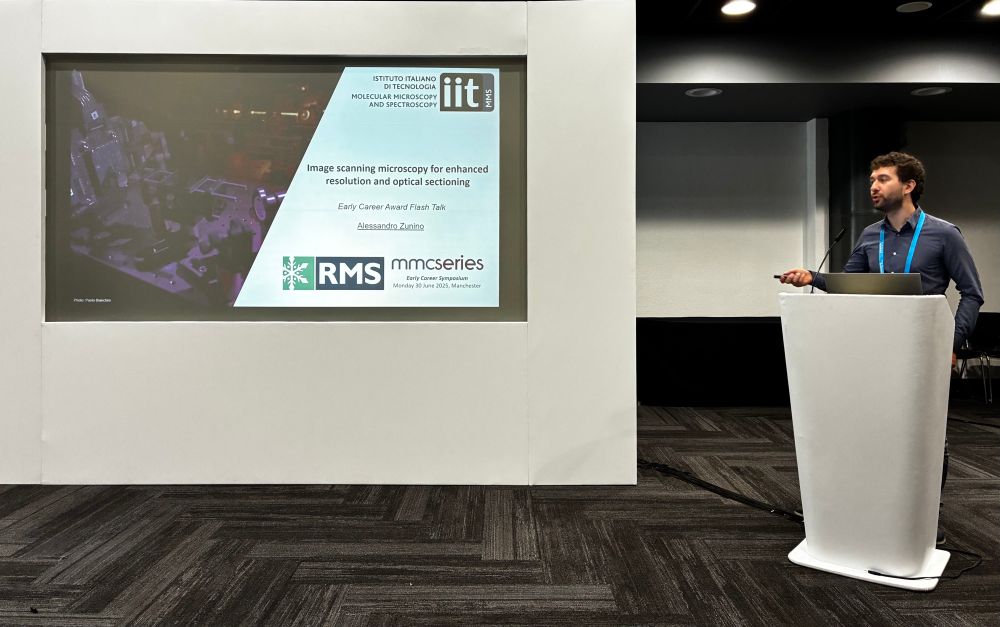
👉 “Photon-Resolved Microscopy : Image-Scanning Microscopy with SPAD Array Detector and Beyond”
👉 youtu.be/Pgn-8F87RUE
@iitalk.bsky.social @cnrsingenierie.bsky.social
#SPADarray #BrightEyes_ERC #photonresolvedmicroscopy #imagescanningmicroscopy
Andrea Bucci and Laurent Le will run two workshops on building a custom #PhotonResolvedMicroscopy system and generating great #ImageScanningMicroscopy images.
Thanks @iitalk.bsky.social @erc.europa.eu @genoainstruments.bsky.social and #PNRR RNA&GeneTheraphy for support. See you at #Mifobio2025

#Mifobio2025
Focus on the module 2 :
👉 the challenges of spatial and temporal quantification
We are delighted to welcome Dr. Giuseppe VICIDOMINI @vicidominilab.bsky.social @iitalk.bsky.social
Lecture - “Photon-Resolved Microscopy : Image-Scanning Microscopy with SPAD Array Detector and Beyond”

Happy to share this article with my views on @elislenders.bsky.social and @vicidominilab.bsky.social work and discussing current developments in single-molecule localization combining structured excitation and detection. So many exciting perspectives in the field!
www.nature.com/articles/s41...

Very happy to be in #ThePhotonics100 list for 2026. Big thanks to everyone in @vicidominilab.bsky.social and to all collaborators for their continuous support!
Interferometric Image Scanning Microscopy for label-free imaging at 120 nm resolution inside live cells https://www.biorxiv.org/content/10.1101/2025.09.19.677416v1

🏆🆕🇪🇺 @elislenders.bsky.social, researcher of the IIT Molecular Microscopy and Spectroscopy unit @vicidominilab.bsky.social , coordinated by Giuseppe Vicidomini, has been awarded an #ERCStG by the @erc.europa.eu
opentalk.iit.it/en/the-resea...

#NewPaper on the blend of computational microscopy with chemical imaging: enabling high-speed quantitative CARS imaging via a new compressive Raman framework.
Kudos @spzhao0724.bsky.social
A collaboration between @lkblab.bsky.social and @ukmlv.bsky.social from Washington Uni St.Louis rdcu.be/eCjLz

Hello! Very excited to share our latest preprint, which is great news for us but terrible news for any diehard fans of PSNR and SSIM as image quality metrics in microscopy... (1/5)
www.biorxiv.org/content/10.1...
and... our program for the upcoming IMAGE SCIENCE🔬 Gordon Research Conference (GRC) is 🎯LIVE🎯. It will take place April 26, 2026 in beautiful tuscany, italy 🇮🇹 🍕 🤌
Please check our stellar line-up, share, and register!
www.grc.org/image-scienc...
We have a FRC module in our Python package: brighteyes-ism.readthedocs.io

"A #SPADarray in any laser-scanning microscope"
Here’s an interview with @zuninoale.bsky.social revealing the behind-the-scenes of our work @iitalk.bsky.social supported by @erc.europa.eu.
#BrightEyes_ERC, #photonresolvedmicroscopy #imagescanningmicroscopy
theanalyticalscientist.com/issues/2025/...

New open access article online: Structured detection for simultaneous super-resolution and optical sectioning in laser scanning microscopy.
go.nature.com/4mfYqb6

A research team led by G. Vicidomini, PI of the IIT Molecular Microscopy and Spectroscopy Lab @vicidominilab.bsky.social , has published in #NaturePhotonics @nature.com a new imaging method that significantly enhances the observation of complex biological tissues.
opentalk.iit.it/en/a-new-mic...
“A #SPADarray in any laser scanning #microscope”. That is the latest effort from a group of people that shared this mantra. Proud of all of them.
@erc.europa.eu
#imagescanningmicroscopy #photonresolvedmicroscopy
#superresolution
#BrightEyes_ERC
doi.org/10.1038/s415...
Traveling Salesman Problem?
#ISMFLUX shows #SPADarray is the future of #MINFLUX, improving localisation robustness and enabling the transformation of a #confocal #microscope into a #MINFLUX system.
www.nature.com/articles/s41...
#photonresolvedmicroscopy #BrightEyes_ERC
@iitalk.bsky.social @erc.europa.eu @ec.europa.eu

Excited to share our paper on ISM-FLUX, our new #MINFLUX-based technology. Achieve sub-10 nm localization uncertainty on single molecules using a straightforward #confocal-based setup with an array detector. 1/6 www.nature.com/articles/s41...
God does not play dice with the universe?
Alessandro Zunino now describes contributions to the Image Scanning Microscope (s^2-ISM) for super-res 3D sectioning much like SIM, and FLIM-capable but with a small confocal photodiode array!

It is time to update our #photonresolvedmicroscope to make it more #smart, and we are looking for a PhD candidate. Here are the application details
vicidominilab.github.io/positions/
If you are interested, please get in touch with me.
@iitalk.bsky.social
#superresolution #microscopy #SPADarray
I smell Richardson-Lucy, here.
Our last paper on s²ISM (super-resolution sectioning #ImageScanningMicroscopy) just got published on Nature Photonics @natphoton.nature.com! Discover how structured detection in laser scanning microscopes enhances both resolution and optical sectioning:
www.nature.com/articles/s41...
🧵1/7

This work was made possible thanks to my amazing colleagues at the @vicidominilab.bsky.social in @iitalk.bsky.social . It is part of the #BrightEyes_ERC project funded by @erc.europa.eu. If you are curious about what else is cooking in the pot, check out our website:
vicidominilab.github.io
🧵7/7

Curious to try it out? You can! Our code is completely free and open source, and our data is available for download. You can find the ready-to-use Python package with examples and documentation here:
github.com/VicidominiLa...
🧵6/7

The benefits of s²ISM extend to any other additional dimension contained in the ISM dataset, such as time and spectrum. We demonstrate superior fluorescence lifetime imaging, enabled by the single-photon timing capabilities of the #SPADarray and strengthened by our approach.
🧵5/7

The concept of s²ISM is general and versatile. It can be applied to any laser scanning technique equipped with a detector array. We demonstrate the feasibility of s²ISM with live-cell imaging, multi-color, and two-photon excitation fluorescence microscopy.
🧵4/7

The ISM dataset is inherently redundant and contains enough information to enable up-sampling of the reconstructed images. s²ISM exploits this property to relax Nyquist’s criterion by a factor of two and enable both optical and digital super-resolution.
🧵3/7

The s²ISM concept is simple: light spreads differently on the detector according to the emitter's axial position. With rigorous physical modelling of ISM image formation, we leverage such unique information to reconstruct only the in-focus fraction of the collected fluorescence photons.
🧵2/7

