Day 5 has an exciting lineup 🤩
@smikulovic.bsky.social talks about helping mice
@nikolaskaralis.bsky.social neuromodulatory circuits
@acnavasolive.bsky.social SWR across species
And me 💁🏽♀️ about theta & movement
#neuroscience
#NeuralMechanismsofCognitiveFunction
sites.google.com/isd.org.br/i...
14.04.2025 13:40
👍 6
🔁 2
💬 1
📌 0

Talk 19:
Next up is @acnavasolive.bsky.social talking about her impressive research on sharp wave ripple analysis across species in health and disease. Andrea is a phenomenal computational neuroscientist 🤩 and I am so excited to hear her speak for the first time!
14.04.2025 17:52
👍 5
🔁 2
💬 1
📌 0
Cont..
@acnavasolive.bsky.social discussing important considerations for identifying hippocampal ripple events in a recording session and how a convolutional NNs might help their identification.
✅
14.04.2025 18:16
👍 2
🔁 1
💬 1
📌 0
This is a great application of our topological analysis of the iEEG waveform space to detect and differentiate interictal discharges, ripples and fast ripples in temporal lobe epilepsy in human. Don’t miss the 📝!! www.biorxiv.org/content/10.1... and the Github 👇🏼
10.03.2025 20:27
👍 10
🔁 1
💬 1
📌 0
It has been a very nice journey of inter-continental collaboration, and hopefully it’s a helpful outcome for the scientific community! If you are interested in these ideas, and want to try it for your own research, please let us know how is it working for you! We are more than happy to discuss! 🤩
10.03.2025 17:34
👍 4
🔁 0
💬 0
📌 0


The manuscript has MANY more interesting analyses. For example, we saw that the intrinsic dimension of IEDs is lower than SWRs; that SWR-IED segregation can depend on the pre-processing; how is coupling between SWRs on macro- & microwires; and the unique spiking modulation patterns in SWRs vs IEDs
10.03.2025 17:34
👍 3
🔁 0
💬 1
📌 0


So based on our current knowledge of rodent and human events, we started by determining what features define SWRs and IEDs, and created a pipeline to analyze epileptic intracranial EEG from start (select the optimal channel) to end (event curation)
10.03.2025 17:34
👍 3
🔁 0
💬 1
📌 0

The motivation of this work comes from the unsolved difficulties of analyzing intracranial EEG in epileptic patients, where the frequent epileptic-like events (interictal epileptiform discharges, or IEDs) interfere with SWR detection, resulting in painfully high false positives🥲
10.03.2025 17:34
👍 3
🔁 0
💬 1
📌 0

2025=(1+2+3+4+5+6+7+8+9)² = 1³+2³+3³+4³+5³+6³+7³+8³+9³. 👉🏼 elpais.com/ciencia/cafe...
04.01.2025 20:02
👍 5
🔁 1
💬 1
📌 0

Membrane potential states gate synaptic consolidation in human neocortical tissue - Nature Communications
Whether and how slow wave activity (SWA) and the underlying membrane potential UP and DOWN states initiate mechanisms that augment memory functions in humans are not fully understood. Here authors use...
More human neurophysiology out today from the Geiger Lab. *Analogue* neuronal output modifies synapses for consolidation during sleep states. With such beautiful data and interpretation it’s easy to forget that the recordings are heroically tough. Fantastic research from @fxmittermaier.bsky.social
12.12.2024 19:57
👍 22
🔁 4
💬 0
📌 1

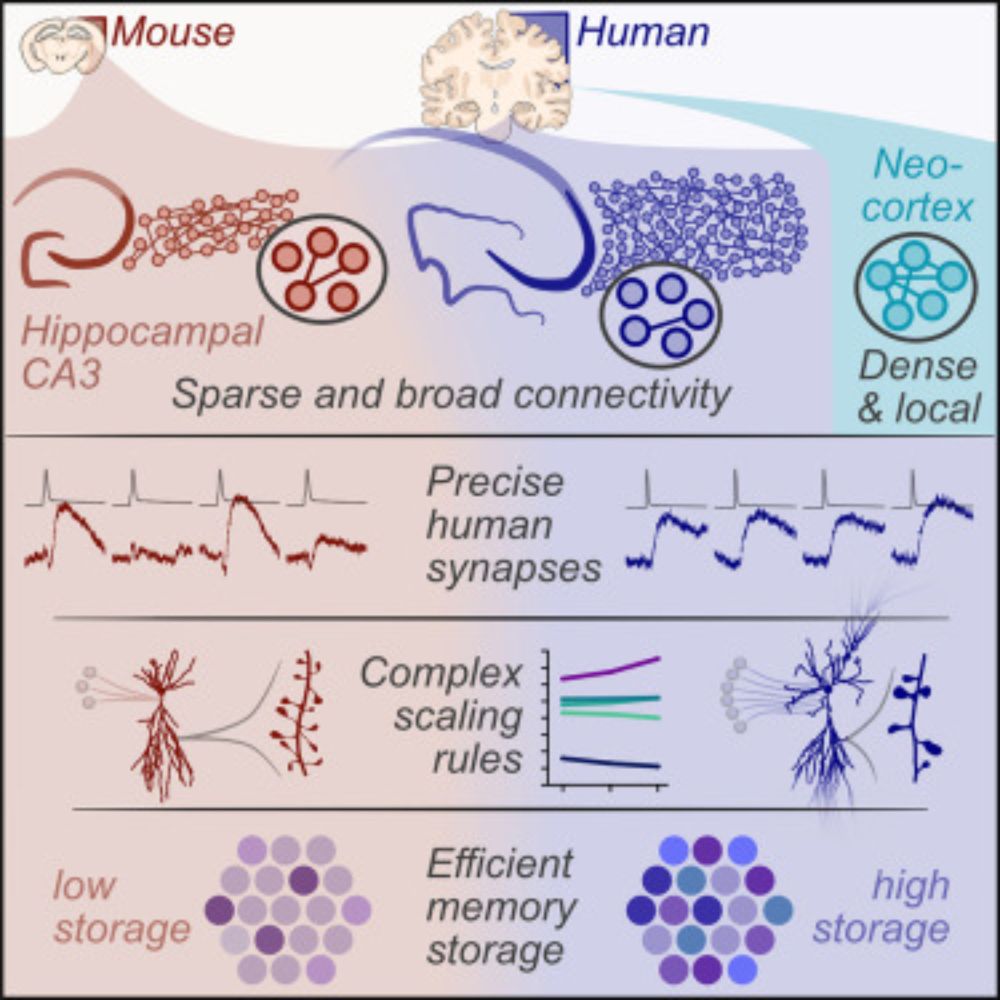
'LICONN' connectomic images of mouse (left) and human (right) CA3, with individual cells segmented (colour). 3D reconstructions of the dendrites show their inputs (lower), with quantification (graph).
With @mojtabart.bsky.social, we applied this to human tissue, and saw that human CA3 cells appear to receive 5 times more DG inputs than mouse cells do! This finding has a lot of potential for powering up our idea of hippocampal computations.
11.12.2024 19:46
👍 5
🔁 1
💬 1
📌 0

Details of CA3 modelling experiment where circuits with the same number of synapses but different circuit architecture were tested for memory capacity. Quantification (right) shows circuit form 'A' has better memory storage (independent of the total size - colours).
Using a Hopfield-like model, @acnavasolive.bsky.social showed that expanding brain size by increasing neuronal number (sparse connectivity) is far better for memory capacity than aiming for dense connectivity (and more inputs per cell). This has some interesting philosophy for brain scaling rules!
11.12.2024 19:46
👍 9
🔁 2
💬 1
📌 0

Human hippocampal slices showing non-sclerotic and sclerotic phenotypes, with cell loss in sclerotic tissue
In a fantastic collaboration with Prof Karl Rössler (MedUniWien), we applied multicell patch-clamp to human hippocampus resected from epilepsy patients. Some samples show sclerosis (disease-led cell loss), but many are perfectly intact. This is the closest to 'wildtype’ human physiology we can get..
11.12.2024 19:46
👍 15
🔁 1
💬 2
📌 0

Example image and recording traces of octuple patch-clamp recording from human hippocampal CA3
We explored CA3, which in theory stores and retrieves memories from interconnected ensembles of pyramidal neurons. With Victor Vargas-Barroso and Rebecca Morse, we looked for these networks using octopatch. From 8 patients and 56 slices we found just 10 connected pairs! (under 1% connectivity)..
11.12.2024 19:46
👍 9
🔁 1
💬 1
📌 0

Measured synaptic connectivity rates between brain areas (neocortex and hippocampus) on the left. Measured synaptic connectivity rates across species in hippocampal CA3 on the right.
To touch base with better characterised circuits, we recorded in neocortex and saw the same dense connectivity that @yangfanpeng.bsky.social, @alleninstitute.bsky.social and others see. In fact CA3 wasn’t just sparse, but gets sparser from mice to humans - opposite scaling to neocortical circuits!
11.12.2024 19:46
👍 12
🔁 1
💬 1
📌 0

Example recordings (left) of CA3 synapses in mouse and human tissue. (Mouse - Red, Human - Blue). Quantification of synaptic potency, reliability, and precision are shown on the right as graphs.
We had the first view on human hippocampal synaptic pairs, and they look slow and integrating as we would expect (perfect for associations!). However they were also much more reliable and precise than seen in rodent research.
11.12.2024 19:46
👍 9
🔁 1
💬 1
📌 0

A collection of images showing the sizes of human and mouse hippocampal slices (grey), an array of CA3 neurons across species (upper) and measured spine densities on the cells (lower), with quantifications.
As you may expect, human neurons were larger than mouse cells, but spine density was a lot lower, so the number of inputs from other neurons in the recurrent network doesn’t change so much. Low spine density and reliable synapses have been seen in other brain areas, so may be human circuit features
11.12.2024 19:46
👍 9
🔁 1
💬 1
📌 0

Schematic of CA3 scaling features (left), and calculation of theoretical connectivity for random recurrent networks with these properties (right).
A bigger difference between human and mouse brains is the number of neurons. This has gone up by about 17 times in CA3! By (very) simple maths, our anatomy data predicts connectivity in a random recurrent network to be pretty much in line with what we record experimentally for CA3 across species.
11.12.2024 19:46
👍 7
🔁 1
💬 1
📌 0

Circuit schematics showing the different connectivity of neocortex (dense local connectivity) and hippocampus (sparse broad connectivity). Dotted square indicates the lens of octuple patch recording to view the circuit.
We think this explains the connectivity and circuit scaling between brain areas - dense local circuits in neocortex, while hippocampal CA3 forms something like one big recurrent network - perfect for associating all the hippocampus’ incoming info. Circuits made to measure!
11.12.2024 19:46
👍 8
🔁 1
💬 1
📌 0

Now that we’re on Bluesky, it’s a good time to bring back #badsciencedrawings – a collection of figures that prove that science is more science than art.
Before Biorender, all we had was MS Paint and a dream. But ovals, lines, and lightning bolts were all we needed
Morales-Botello et al., 2012
04.12.2024 20:55
👍 159
🔁 45
💬 5
📌 9
So nice to see science sparking again 🦋 Hello everyone!
19.11.2024 21:15
👍 6
🔁 0
💬 0
📌 0

This paper is incredible. EM level connectomics on a light microscope.
www.biorxiv.org/content/10.1...
14.11.2024 01:22
👍 123
🔁 28
💬 3
📌 4
Following the great migration! Leading the @lmprida.bsky.social lab. Hippocampal circuits, oscillations and memory.
14.11.2024 06:40
👍 43
🔁 2
💬 1
📌 0
go.bsky.app/LdtUYZS
I created a starter pack for the growing community of hippocampus physiologists that have joined the great migration. Also included the physiology-adjacent.
Tell me who I haven’t found yet
12.11.2024 21:00
👍 54
🔁 27
💬 16
📌 2