Registration and abstract submission link below:
www.sggd2026.no/abstract-sub...
02.03.2026 10:41
👍 0
🔁 0
💬 0
📌 0

@cheralab
We are using hiPSC-derived organoids, targeted cell ablation, genetic cell tracing, gene expression tuning and multiomics to study regulators of cell specification decisions and cell plasticity. 📍 UiB Norway 👩🏻🎓 UniGe Switzerland https://chera.w.uib.no/
Registration and abstract submission link below:
www.sggd2026.no/abstract-sub...


Last day to send an abstract for the:
10th Meeting of the Study Group on Genetics of #Diabetes
#SGGD which will take place at the Solstrand Hotel in the magnificent landscape by the Bjørnafjord, just 30 km from the center of in Bergen, Norway.

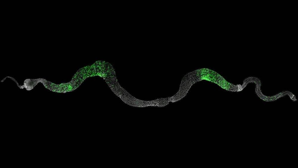
#FluorescenceFriday
#devbio
pic credit: Ulrik Larsen
… qRT-PCR 😀 not T
I used once proteomics to validate NGS. It was called "creative" but still not an qRT-PCT…
I'm with you on this! 💯
⬇️

Congratulations 🎉 to @unger-lucas.bsky.social on successfully defending his PhD with the thesis “Dissecting HNF1A’s Role in Early Cell Fate Decisions and Their Impact on Pancreatic Islet”! 👏 🎓
#cellfate #devbio

Professor Emerita Birgitta Åsjø @unibergen.bsky.social is introducing Lucas @unger-lucas.bsky.social as he starts his PhD work presentation focused on the Dissecting HNF1A’s Role in Early #CellFate Decisions & their Impact on Pancreatic #Islet
@cheralab.bsky.social
#PhDLife #PhDdefense #devbio

The Elbe Program is offering positions for independent postdocs at the interface between theory (physics, maths, computing) and biology. Join us in Dresden! Deadline 13 March.

@unger-lucas.bsky.social will defend his PhD work 🎓@unibergen.bsky.social
🕒Tuesday, 24th of February at 12.15
📍Auditorium 2, BB-bygget, Jonas Lies vei 91
Trial Lecture at 10.15
“Transcriptional regulation in the developing pancreas: From genome architecture to phenotypes”
#UiB 🇳🇴 #devbio #PhDLife

🧪 Come work with us in Bergen! 🌊
Do you have a good understanding of paleoceanography & a masters degree in Geosciences, Marine Biology or similar - this might be your chance to become a researcher 💥
@i2b-erc.bsky.social
@polhavet2050.bsky.social
Read more and apply here: lnkd.in/e8KBAyPi

Promotional graphic announcing “Nominations for EASD Prizes 2026.” The left side features a dark blue background with the headline “Honouring excellence in diabetes research!” and text inviting nominations to recognise outstanding colleagues. The right side shows abstract blue light rays and curved white and light-blue graphic rings. The EASD logo appears at the bottom left, with the website link “easd.org/prizes” at the bottom right.
⌛ Few days left to #nominate a colleague who’s made an outstanding contribution to #diabetes #research & #care. Each year, EASD honours pioneers & leaders with 6 prestigious #prizes.
📅 Deadline: 15 Feb 2026 | 🕛 12:00 CET
Spread the word & nominate now 👉 www.easd.org/prizes/

Webinar alert
Learn how to generate 3D human multi-cell type neurospheres from iPSCs without necrotic core formation.
Featuring Stefan Wendt, PhD (UBC)
Date: March 25 | Time: 12:00 pm ET
Free for all
Register here: https://www.ascb.org/ascb-meetings/3d-human-multi-cell-type-meurospheres-from-ipscs/

John Gurdon taught us that the beginning of life is never truly lost — only waiting to be reawakened. Long before his ideas were accepted, he showed that a cell remembers more than it appears to know: that within a differentiated nucleus lies the latent capacity to begin anew go.nature.com/3Mf8SD0
Rather proud that our mouse got its own official name 🐀
thanks @mousegenome.bsky.social
www.informatics.jax.org/allele/MGI:8...

Meet Skatt, my trusted grant advisor.
He has strong opinions. Mostly about who's the Chair Master.
#JellyBellyFriday
Professor Ville Hietakangas calls for patience from research funders: the time frame in basic research is long, and financial concerns undermine creativity. Read more in our article: sigridjuselius.fi/en/news/scie...
We are recruiting a postdoc to join a Wellcome Trust-funded project at LSI-Exeter, investigating the role of neural-pancreas dynamics in maintaining glucose homeostasis. 🐟🔬🧠
@lsiexeter.bsky.social #zebrafish #islet #postdocjobs
jobs.exeter.ac.uk/hrpr_webrecr...
Please repost 🙏

Studies in cells and mice reveal that the human GLP1R A316T variant exhibits characteristics of constitutive activation but dampened GLP-1RA responses www.science.org/doi/10.1126/...


Just discovered the wonderful covers of 'Genes to Cells', the journal of the Molecular Biology Society of Japan @mbsj-official.bsky.social – absolutely beautiful!
here some examples inspired by mitosis, CRISPR, the DNA helix, and plant pigments

Promotional graphic announcing that scientific submissions for EASD 2026 are open. Text highlights the dates “28 September-02 October” and the location “Milan, Italy.” A message invites researchers to submit abstracts to share their work with the global diabetes community. The design features red and white brush-stroke panels and a stylised skyline of Milan on the right, with “EASD 2026 Milan” and “easd.org” at the bottom.
🎯 Started preparing your #abstract submission for the 62nd EASD Annual Meeting? This is your sign to get started & share your #diabetes #research with the global community in Milan 🇮🇹 from 28 Sept - 02 Oct 2026.
🗓️ Deadline: 1 Apr 2026 | 🕛 18:00 CEST
Submit via MyEASD 👉 www.easd.org/annual-meeti...

Call for Applications graphic for the EFSD Rising Star Fellowship Programme and EASD Rising Star Symposium. Text encourages young researchers in Europe to develop innovative diabetes research. Supported by Novo Nordisk through a donation. Includes EFSD logo, website europeandiabetesfoundation.org, abstract blue and green shapes, and an image of a researcher using a microscope.
📢🚨 Don’t miss this: Apply for the #EASD Rising Star Symposium and #EFSD Rising Star Fellowship Programme!
Eligibility: EASD members, within 7 years of PhD/MSc/MD
⏰ Deadline: 17 Feb 2026 | 12:00 CET
👉 Apply: www.europeandiabetesfoundation.org/programmes/p...
Supported by Novo Nordisk
#diabetes

Would you like to participate in the peer review process at Communications Biology? Please fill out this form to express your interest to act as a reviewer📝 We particularly encourage early-career researchers and applicants from underrepresented groups in research. forms.office.com/e/fv22f9aRt5

Fig. 1. Animal cap assay and sandwich method as in vitro induction systems. In amphibians, a blastocoel cavity clearly forms inside the animal hemisphere during the blastula and early gastrula stages. The cap-like portion lining the roof of the blastocoel cavity is the animal cap. This region consists of a sheet of pluripotent cells, organized into one or several layers. In the animal cap assay, the animal cap was treated with a physiological saline solution containing inducing factors and then cultured. Depending on the type, concentration, and duration of exposure to the inducing factors, animal caps can differentiate into various cell types. In contrast, the sandwich method, involves culturing the inducer source in between two animal caps. In this technique, the sources of induction can include the dorsal lip of the blastopore (organizer), adult tissues, pelletized soluble factors, or animal caps pretreated with soluble factors. In this figure, activin is used as an example of an inducing factor.
![Fig. 12. Summary of the in vitro induction system using activin as an inducing factor.
This in vitro induction system utilizes activin and retinoic acid as inducing factors to treat animal caps, employing techniques such as animal cap assay, dissociation/reaggregation protocol, and the sandwich method. By applying these methods, various levels of self-organization can be replicated and controlled in vitro, ranging from lower-order cell differentiation to higher-order tissue differentiation, organogenesis, and even the formation of fundamental body plans. Abbreviations: Dorsal [D], ventral [V], and retinoic acid [RA].](https://cdn.bsky.app/img/feed_thumbnail/plain/did:plc:cnykzbo2impysrf6a6scy6oh/bafkreiarcsezsnfoj4xmlow6thcxsfy6wsfxfujek53lgt6wsw2k2wshqe@jpeg)
Fig. 12. Summary of the in vitro induction system using activin as an inducing factor. This in vitro induction system utilizes activin and retinoic acid as inducing factors to treat animal caps, employing techniques such as animal cap assay, dissociation/reaggregation protocol, and the sandwich method. By applying these methods, various levels of self-organization can be replicated and controlled in vitro, ranging from lower-order cell differentiation to higher-order tissue differentiation, organogenesis, and even the formation of fundamental body plans. Abbreviations: Dorsal [D], ventral [V], and retinoic acid [RA].
![Fig. 11. Formation of embryoids by artificial activin concentration gradients.
To create embryoids, animal caps were prepared through treatment with low (0.5–1 ng/ml), intermediate (5–10 ng/ml), or high (50–100 ng/ml) concentrations of activin. These three types of activin-treated animal caps were then sequentially arranged and cultured with untreated animal caps. After 3 days of culture, embryoids with distinct head and trunk-tail structures were formed (A). Histological sections revealed differentiation into head tissues, such as the cement gland [cg] and eyes, and trunk-tail tissues including the ear vesicle [ev], brain [br], notochord [not], muscle [mus], and gut (B). When newt embryos are used in similar combination cultures, neural plate structures forming the brain [white arrow] and axial structures forming the trunk-tail regions [black arrow] are sometimes observed (C).](https://cdn.bsky.app/img/feed_thumbnail/plain/did:plc:cnykzbo2impysrf6a6scy6oh/bafkreig36tdo3ewakfxhn66uzs2kfqlkjjoebdcfh5rzy3ubxkfnn7t5iu@jpeg)
Fig. 11. Formation of embryoids by artificial activin concentration gradients. To create embryoids, animal caps were prepared through treatment with low (0.5–1 ng/ml), intermediate (5–10 ng/ml), or high (50–100 ng/ml) concentrations of activin. These three types of activin-treated animal caps were then sequentially arranged and cultured with untreated animal caps. After 3 days of culture, embryoids with distinct head and trunk-tail structures were formed (A). Histological sections revealed differentiation into head tissues, such as the cement gland [cg] and eyes, and trunk-tail tissues including the ear vesicle [ev], brain [br], notochord [not], muscle [mus], and gut (B). When newt embryos are used in similar combination cultures, neural plate structures forming the brain [white arrow] and axial structures forming the trunk-tail regions [black arrow] are sometimes observed (C).
![Fig. 7. In vitro heart formation and in vivo transplantation experiment.
When treated with a high concentration of activin, the animal caps of Xenopus embryos did not differentiate into heart tissue. However, if the animal cap dissociates into individual cells before activin treatment and then reaggregates, it forms a beating heart [arrow] with 100 % efficiency (A). This heart expresses differentiation marker genes, such as Nkx2.5, GATA-4, Tbx5, MHCα, TnIc (cardiac troponin I), and ANF, none of which are expressed in an animal cap treated with activin alone, without dissociation/reaggregation (B). Electron microscopy reveals the presence of intercalated discs [id] specific to the cardiac muscle, along with visible mitochondria [m] and Z-bands [z] (C). When the reaggregated heart tissue is orthotopically transplanted into the cardiac primordium of a neurula-stage embryo, it integrates without rejection and continues to beat (D), although it does not persist through host metamorphosis. In contrast, when the reaggregated tissue is ectopically transplanted into the ventral region of the neurula, it begins to beat synchronously with the host heart and gradually reddens as it initiates blood circulation (E).](https://cdn.bsky.app/img/feed_thumbnail/plain/did:plc:cnykzbo2impysrf6a6scy6oh/bafkreig77lwzs67jdcjwi2gjsany3shyoa5vj2dpetburc3pegwxeb45a4@jpeg)
Fig. 7. In vitro heart formation and in vivo transplantation experiment. When treated with a high concentration of activin, the animal caps of Xenopus embryos did not differentiate into heart tissue. However, if the animal cap dissociates into individual cells before activin treatment and then reaggregates, it forms a beating heart [arrow] with 100 % efficiency (A). This heart expresses differentiation marker genes, such as Nkx2.5, GATA-4, Tbx5, MHCα, TnIc (cardiac troponin I), and ANF, none of which are expressed in an animal cap treated with activin alone, without dissociation/reaggregation (B). Electron microscopy reveals the presence of intercalated discs [id] specific to the cardiac muscle, along with visible mitochondria [m] and Z-bands [z] (C). When the reaggregated heart tissue is orthotopically transplanted into the cardiac primordium of a neurula-stage embryo, it integrates without rejection and continues to beat (D), although it does not persist through host metamorphosis. In contrast, when the reaggregated tissue is ectopically transplanted into the ventral region of the neurula, it begins to beat synchronously with the host heart and gradually reddens as it initiates blood circulation (E).
A fascinating review on the role of Activin in organ induction. Isn't it wild that in Xenopus embryos, a piece of the animal cap can be induced with Activin at different concentrations and buffers to form the ❤️, kidney, the pancreas, head, tail, and even a whole embryoid 🤯:
doi.org/10.1016/j.cd...

Explore emerging research with field leaders Keystone Symposia #IsletBiology & #Diabetes : #BetaCell Compensation, Failure & Recovery, this March in Breckenridge! #KSDiabetes26
For more info:
➡️ https://hubs.la/Q03X7Ckz0

That 5th sample...
#lama #portal #aperturesciencesentryturret #thinkingwithportals


#Norway at its best. From my backyard #northernlights #aurora

Piece on why Nature became so influential and “important”. This quote: “history shows that much of its success has been circumstantial and opportunistic rather than meritorious…we should not mistake the system that emerged for one that reliably surfaces the best work.” www.asimov.press/p/nature

well, Nicolae Paulescu isolated insulin, although he called it pancreine and even try to patent it in 1916 - but Banting, Best and Macleod got it to work in humans. Probably it matters who gets the best outcome of research more than who had the idea first…
en.wikipedia.org/wiki/Nicolae...
Not sure, it takes a lot to keep up with being scientist, but I would say that some people truly dislike other scientists…