#ImageOfTheMonth March 2026
Dividing Arabidopsis epidermal cells imaged by confocal microscopy after expansion. Microtubules guided cell plate formation revealed in 3D with depth-code.
Image: Magali Grison @lbm-bordeaux.bsky.social @gtexm.bsky.social
#Microscopy #PlantScience #CellBiology
02.03.2026 14:36
👍 26
🔁 10
💬 0
📌 0

Save the date 🤩🤩🤩!
Our student-led Young Scientist Symposium will take place on the 4th & 5th of June this summer!
More information and registration available soon!
29.01.2026 12:03
👍 7
🔁 8
💬 0
📌 0

🏆 Image of the Year 2025
226 votes, 12 Images of the Month, and a race decided by just one vote 😮
With 58 votes, @tdhellemmes.bsky.social wins for his cleared brain image acquired using light-sheet microscopy and will proudly carry the colors of the BIC 🎉🔬✨
02.02.2026 15:14
👍 9
🔁 3
💬 0
📌 1

#ImageOfTheMonth February 2026 ✨
Fluorescent fibers (hundreds of µm) of a Platinium complex visualized in Fiji using orientation-based pseudocolors 🎨. Image acquired during the BIC hands-on microscopy training 🔬
Credits: F. R. Lauro, A. Virepinte (ISM), J. Teillon (BIC)
02.02.2026 15:05
👍 6
🔁 3
💬 0
📌 0

[WEBINAR] Join the next France Volume-EM WebCafe!
Karin Pernet-Gallay from the GIN (Grenoble) will present her latest project, revealing new insights into astrocyte structure & function. These results would never have been obtained without 3D EM!
🗓️ January, 29th - 13:00
🔗 Join here: bit.ly/49Ux45l
26.01.2026 10:42
👍 1
🔁 1
💬 1
📌 0
✨ En ce début d’année, l’équipe du pôle photonique s’est dit qu’il était temps de changer des slides et des listes d’équipements 📄❌
👉 Résultat : un mini-film un peu décalé pour présenter nos systèmes de microscopie, d’optique et d’instrumentation 🔬
La photonique, mais en version musicale 🎶
23.01.2026 14:45
👍 5
🔁 2
💬 1
📌 2

A colorful start to 2026! 🌿 #ImageOfTheMonth
M. Batsale (Plant Virology team, @inrae-bfp.bsky.social ) captured a Nicotiana benthamiana cell infected with turnip mosaic virus — peroxisomes (yellow), viral vesicles (magenta) & chloroplasts (blue).
02.01.2026 09:58
👍 16
🔁 7
💬 0
📌 0

✨ December 2025 #ImageOfTheMonth ✨
Whole rat hearts cleared with iDISCO reveal sympathetic innervation (orange) and vascularization (green). 💛💚
Work by S. Bertrand & A. Bonilla (INCIA) with S. Martin & I. Brunet (CIRB), imaged by J. Teillon (BIC).
#Microscopy #iDISCO #BICBordeaux
04.12.2025 11:30
👍 16
🔁 5
💬 0
📌 0

November #ImageOfTheMonth goes to M.Pujol from LPCO lab (UMR 5629). Acquired with Transmission Electron Microscopy, it shows a polystyrene (PS) latex obtained by post-dispersion of a yogurt cup made of PS in water using a surfactant. The resulting particles are highly heterogeneous and large.
03.11.2025 11:03
👍 4
🔁 1
💬 0
📌 0

October #ImageOfTheMonth is from O. Barba-Vila from @iins-bordeaux.bsky.social Slice of the gustatory cortex showing axons from the gustatory thalamus (G), basolateral amygdala (R) and a thick-tufted layer 5 neuron (W), recorded through patch clamp and imaged via biocytin-streptavidin labelling
07.10.2025 07:48
👍 6
🔁 1
💬 0
📌 0

September #ImageOfTheMonth is from @rkinet.bsky.social from @imn-bordeaux.bsky.social
This is a maximum projection of a confocal image of a microglia cell (blue) and its nucleus (red), internalising multiple nanoparticles (yellow), one day after supranigral injection in C57B6-J mouse
08.09.2025 09:09
👍 8
🔁 3
💬 0
📌 0

August #ImageOfTheMonth is from L.Congiu from
@imn-bordeaux.bsky.social. This is a 3D reconstruction of a CD68-positive particles (white) within the cell body of microglia cells (Red) in the mouse cortex. The confocal Z-stack was processed with Imaris software.
07.08.2025 18:56
👍 4
🔁 2
💬 0
📌 0
It’s good to be back at the BIC! @bic-bordeaux.bsky.social 🔬
23.07.2025 15:23
👍 7
🔁 1
💬 0
📌 0

July #ImageOfTheMonth is from A.Drouet from
@iins-bordeaux.bsky.social. This image represents a neurosphere (150x150 µm) formed from dissociated hippocampal neurons cultured for 14 days under non-adhesive conditions. Yellow shows GFP expressing neurons and nuclei are showed in cyan.
04.07.2025 09:36
👍 5
🔁 3
💬 0
📌 1
@jeremie-bic.bsky.social
17.06.2025 12:53
👍 2
🔁 0
💬 0
📌 0

June #ImageOfTheMonth is from L.Boutaleb from @inrae-bfp.bsky.social. They study the impact of heat stress on cell division and DNA replication on the tomato meristem. On this confocal image, EdU staining (magenta) detects replicating DNA and Hoechst labeling (cyan) shows all cell nuclei.
05.06.2025 08:49
👍 6
🔁 2
💬 0
📌 0

May #ImageOfTheMonth was from C.Mazocco & M.Darricau from @imn-bordeaux.bsky.social. Staining on spinal cord slices reveal TDP43 protein in the nucleus of neurons (Red). Motoneurons and synaptic terminations were stained targetting ChAT (Cyan).
05.06.2025 08:44
👍 7
🔁 4
💬 0
📌 0
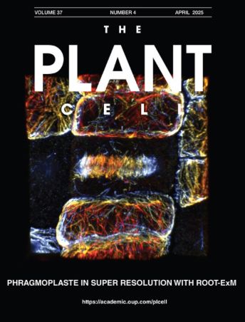
Time to expand your vision! 👀🌱
Our latest work on ExM in plants is out
Back-to-back with the Danzl 's lab (@ISTAustria) led by Magali Grison @lbm-bordeaux.bsky.social with @monica-bic.bsky.social and @gmaucort.bsky.social from the @bic-bordeaux.bsky.social .
academic.oup.com/plcell/artic...
17.04.2025 08:49
👍 41
🔁 25
💬 1
📌 1

Root expansion microscopy: A robust method for super resolution imaging in Arabidopsis
Root expansion microscopy (ROOT-ExM) achieves super-resolution expansion microscopy in plants.
Finally out!, after a few months of delay, our protocol on ExM in plant roots is now available in The Plant Cell
academic.oup.com/plcell/artic...
Beautiful images in a great collaboration of the @bic-bordeaux.bsky.social with @emmanuellebayer.bsky.social and Magali Grison @lbm-bordeaux.bsky.social
14.04.2025 09:47
👍 35
🔁 12
💬 1
📌 0

April #ImageOfTheMonth is from H. Vaïtinadapoulé (BiiO EA 2521). Here is a guttae, a “drop-like deposits” of extracellular matrix components on the surface of the cornea. This image was acquired using scanning electron microscopy with the help of F.Decoeur from the EM unit of the BIC.
01.04.2025 16:19
👍 9
🔁 2
💬 0
📌 0

March #ImageOfTheMonth goes to M.Bonhivers from MFP lab (UMR 5234).
Trypanosoma brucei cell was extracted in detergent, expanded 4.2 fold using U-ExM, labelled for the microtubule cytoskeleton (magenta) and for the Hook Complex (yellow), and images usinf scanning confocal microscopy.
03.03.2025 11:00
👍 19
🔁 5
💬 0
📌 1

Super-resolution microscopy unravels a dynamic tubular network that is stabilized by pre-existing Golgi and lipids to mature into new Golgi pre-cisternae, @bic-bordeaux.bsky.social, @cnrsbiologie.bsky.social, @cnrsaquitaine.bsky.social, #Lipids, #ExpansionMicroscopy, #PlantScience, #PlantCellBiology
25.02.2025 10:11
👍 9
🔁 2
💬 1
📌 1
2025 Speakers – International Frontiers in Neurophotonics Symposium
Save the date !
Frontiers in Neurophotonics Symposium 2025 in Bordeaux
October 6-9
frontiersneurophotonics.org/2025-speakers/
🧪
05.02.2025 14:08
👍 10
🔁 4
💬 0
📌 2
Top pictures display
dorso-to-ventral samples of bottom max projection (from left to right).
03.02.2025 17:01
👍 0
🔁 0
💬 0
📌 0

February #ImageOfTheMonth is from
@tdhellemmes.bsky.social
. GFP expressing Relaxin3 neurons from the nucleus incertus and their projections are followed through the whole mouse brain using Adipoclear+ method and Light Sheet microscopy. Scale:1mm
03.02.2025 17:00
👍 8
🔁 3
💬 2
📌 2

Happy new year! Let's start 2025 with an #ImageOfTheMonth from N. Bollier @inrae-bfp.bsky.social. Scanning confocal 3D reconstruction of a tomato shoot apical meristem. The fixed meristem was cleared using clearsee and the cell walls are stained by calcofluor white.
05.01.2025 15:39
👍 21
🔁 8
💬 1
📌 1

Kaleidoscopic view of my tracing experiment, from top to bottom.
Our great achievement with @jeremie-bic.bsky.social in @bic-bordeaux.bsky.social for @imn-bordeaux.bsky.social
30.12.2024 16:27
👍 6
🔁 2
💬 0
📌 0


Icing stars and flowers in electron microscopy.
With the courtesy of Isabelle Svahn in @neurobordeaux.bsky.social and @bic-bordeaux.bsky.social
29.12.2024 14:56
👍 7
🔁 2
💬 0
📌 0

Hello, the BIC is starting its Bluesky experience with its December #ImageOfTheMonth. Beautiful rat neurons imaged in STED 😍 by T.Cloâtre from @iins-bordeaux.bsky.social !
12.12.2024 10:30
👍 30
🔁 3
💬 0
📌 0
For Christmas this year the BIC asked for a new 🔬booking website and Santa heard us 😊. So now we are asking our users to help us find it the best name. If you have ideas and want to participate please help yourself:
forms.gle/GBJ3zymegDSr8p…
23.01.2023 11:13
👍 0
🔁 0
💬 0
📌 0