Double PI act @sorengrubb.bsky.social @amreenmughal.bsky.social continuing in brains with #cilia & #centrosome diversity across microvascular system, with striking species differences on cilia metrics & directionality of spaces sampled. Deep read: www.biorxiv.org/content/10.1... #UKCilia 5/8
13.01.2026 23:00
👍 3
🔁 1
💬 1
📌 0
Want to know about our latest research on #primarycilia and #centrosomes in #brain #microvasculature? Join this e-symposium tomorrow! 🧪
12.01.2026 20:16
👍 13
🔁 1
💬 0
📌 0
Thanks for a great 2025!🎆🥳🎉
We’re proud to share highlights from this year at Neuroscience Academy Denmark (NAD) – showcasing the incredible work of our PhD students. 👩🎓👨🎓
A big thank you to @lundbeckfonden.bsky.social for supporting our mission. 🙏
#Neuroscience #PhD #Research #Collaboration 🧪
16.12.2025 12:07
👍 10
🔁 2
💬 0
📌 0
Also thanks to @helenelab.bsky.social for access to the CA1 dataset!
20.11.2025 21:48
👍 2
🔁 0
💬 0
📌 0

a man in a suit and tie is giving a thank you sign .
ALT: a man in a suit and tie is giving a thank you sign .
We would like to acknowledge the amazing #OpenScience #ElectronMicroscopy datasets by @alleninstitute.org , @hhmijanelia.bsky.social and other sources!
This work is a collaboration between me at @ucph.bsky.social, @amreenmughal.bsky.social and Vanshika at #NIH, and Carolyn and Jennifer at #Janelia
20.11.2025 21:03
👍 6
🔁 0
💬 1
📌 0

Graphical abstract showing gradient distribution of primary cilia in the mural cells. Primary cilium frequency and lengths are reversely proportional to smooth-muscle actin expression.
🧩 Summary:
✅ #Pericytes have #PrimaryCilia with an arterio-venular gradient🏳️🌈
✅ 🚶♀️🧠 ECs have primary #cilia, 🐁🧠 ECs don’t
✅ EC polarity aligns opposite to #BloodFlow 🧭
❔Lots of open questions about cilia’s role in #NeurovascularCoupling!
Read the full preprint: 🔗 www.biorxiv.org/content/10.1...
🧪🧵8/8
20.11.2025 21:03
👍 11
🔁 1
💬 1
📌 0
🧭 #EndothelialCells are polarized against #BloodFlow direction ⤴️. #Centrioles are consistently upstream of the #nucleus. This polarity persists even in fixed tissue and could serve as a structural marker for flow direction. 📌
🧪🧵7/8
20.11.2025 21:03
👍 6
🔁 0
💬 1
📌 0

A schematic illustration of the neurovascular unit highlighting primary cilia in ECs.
😮Surprise: 🚶♀️🧠 #EndothelialCells (EC) have #PrimaryCilia facing the vessel lumen, but 🐁🧠 ECs don’t. ~18% of 🚶♀️🧠 ECs are ciliated, mostly in arteriolar-end capillaries. What do these #cilia sense in the 🩸?
🧪🧵6/8
20.11.2025 21:03
👍 4
🔁 0
💬 1
📌 0
In 🐁, some #pericyte #PrimaryCilia penetrate #astrocytic #endfeet and reach the #neuropil - sometimes near #synapses! Could these #cilia act as sensory “antennae” 📡for #NeurovascularCoupling?
🧪🧵5/8
20.11.2025 21:03
👍 3
🔁 0
💬 1
📌 0
In 🐁🧠, #ultrastructure shows that #pericyte #cilia are often ensheathed by #astrocytic #endfeet. In 🚶♀️🧠, #astrocytes are swollen and cilia stay confined within the basal lamina. What are they sensing from the astrocytes?
🧪🧵4/8
20.11.2025 21:03
👍 5
🔁 1
💬 1
📌 0
🗺️We mapped #MuralCells — including #pericytes and #smoothmuscle — as vectors➡️ to visualize where #PrimaryCilia🌟 occur across the 🐁🧠 #microvasculature.
The result? A striking pattern: #cilia are abundant on #capillaries and #venules, but almost absent on #arterioles.
🧪🧵3/8
20.11.2025 21:03
👍 3
🔁 1
💬 1
📌 0

(d) Percent distribution of primary cilia in MCs after microvascular stratification into pial and parenchymal vessels, including 1-3rd order capillaries from the MICrONS (mouse visual cortex), CA1 (mouse hippocampus), and h01 (human cortex) datasets. Note that neither CA1 nor h01 contain any pial vessels. (e) Lengths of MC primary cilia in pial and parenchymal vessels, including 1-3rd order capillaries MCs from MICrONS.
📊 #Pericytes are frequently ciliated in both 🐁 and 🚶♀️🧠—but mostly near #venules and venous-end #capillaries.
Venular mural cells: up to 90% ciliated in 🐁 datasets!
Arteriolar mural cells: almost none.
Why this gradient? Flow, pressure, permeability?
🧪🧵2/8
20.11.2025 21:03
👍 1
🔁 0
💬 1
📌 0
New on bioRxiv! 🚨
“Pericyte and Endothelial Primary Cilia and Centrioles have Disparate Organization Across the Brain Microvasculature”
This project studying #microvascular cells took an unexpected turn when we discovered that #pericytes have primary #cilia!
🔗 www.biorxiv.org/content/10.1...
🧪 🧵1/8
20.11.2025 21:03
👍 38
🔁 11
💬 1
📌 2
@martienkas.bsky.social @brittae.bsky.social @dresler.bsky.social
14.11.2025 13:03
👍 0
🔁 0
💬 0
📌 0
🎥 NAD 2025 Annual Meeting Highlights
From Oct 3–5, Neuroscience Academy Denmark hosted:
✅ 400+ participants
✅ 100 posters
✅ Keynotes on precision psychiatry, brain barriers, ultrasound treatments, brain stimulation, sleep & circadian rhythms
We look forward to next year!
#Neuroscience #NAD2025 🧪
14.11.2025 12:58
👍 5
🔁 1
💬 1
📌 0

That reminds me of Thor fighting “midgårdsormen” the world serpent 🤣
27.09.2025 08:31
👍 2
🔁 0
💬 0
📌 0
“Well I don’t like that study because it doesn't give me the result I want’
08.09.2025 14:32
👍 139
🔁 56
💬 7
📌 7

We are excited to report:
Cerebral blood flow is modulated by astrocytic cAMP elevation independently of IP3R2-mediated Ca2+ signaling in mice
Vittani et al. PNAS (2025)
pnas.org/doi/10.1073/...
02.07.2025 12:13
👍 29
🔁 12
💬 2
📌 0

The first Master’s student with me as main supervisor graduated today! I’m proud 🥹
24.06.2025 17:37
👍 13
🔁 0
💬 0
📌 0
Thanks Amreen! ❤️
10.06.2025 20:14
👍 1
🔁 0
💬 0
📌 0
In older mice, where #MyogenicTone is weaker, #PrecapillarySphincters become even more critical barriers against #BloodPressure surges—compensating for #VascularStiffening and remodeling.
10.06.2025 19:38
👍 2
🔁 0
💬 1
📌 0
These changes suggest that #PrecapillarySphincters protect the capillaries 🛡️ by acting as a dynamic #resistance that regulates #capillary #BloodFlow and #BloodPressure.
10.06.2025 19:38
👍 1
🔁 0
💬 1
📌 0
Using #InVivo #VolumeImaging, we found #age related #vascular changes:
• ↑ #Tortuosity of #PenetratingArterioles
• ↓ #Arteriolar-capillary density
• ↑ #Capillaries at the venous end + #aneurysm-like bulbs downstream of #PrecapillarySphincters.
10.06.2025 19:38
👍 0
🔁 0
💬 1
📌 0
By recording #cortical #angiograms during i.v. injection of FITC-dextran 🟢 in #adult and #old #NG2DsRed mice, we time-colored the #arterioles 🔴 and #venules 🔵. We found that #OldMice have less #collaterals and more #tortuous #PenetratingArterioles.
10.06.2025 19:38
👍 0
🔁 0
💬 1
📌 0

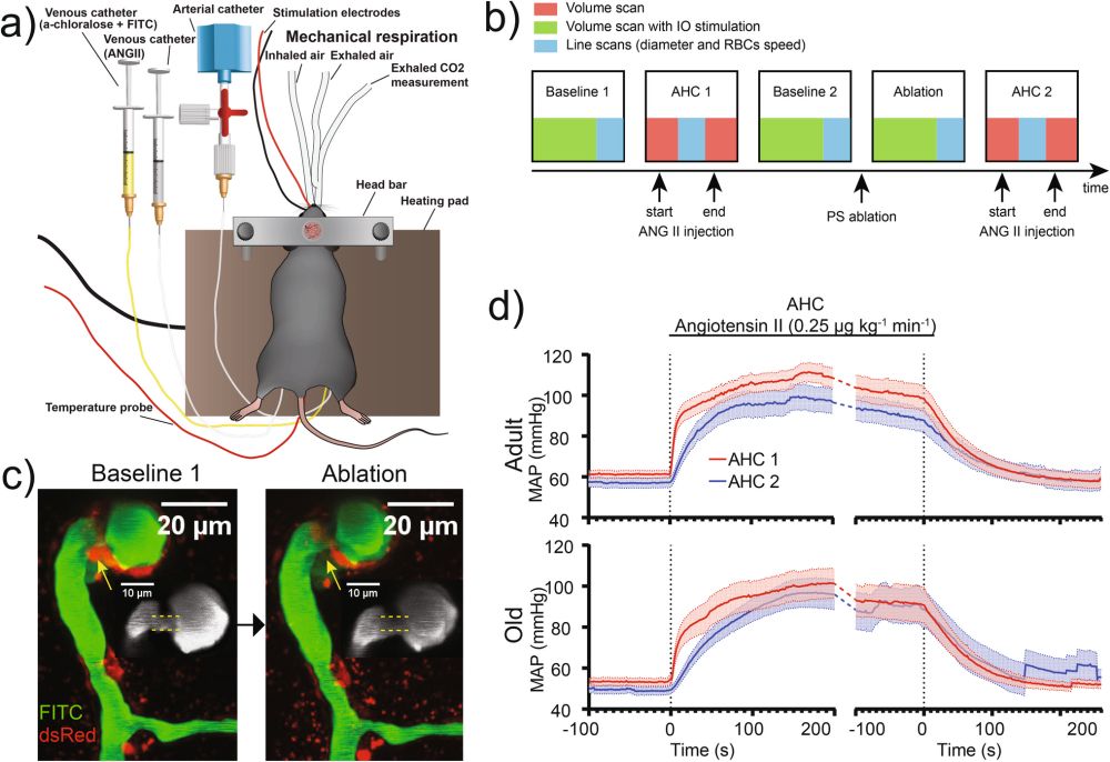
Figure showing how we used the two-photon laser to ablate the pericyte around the precapillary sphincter.
After #LaserAblating pericytes around #PrecapillarySphincters, we observed impaired #MyogenicResponse and #NeurovascularCoupling—demonstrating #PrecapillarySphincter pericyte contractility is essential.
10.06.2025 19:38
👍 0
🔁 0
💬 1
📌 0

Figure showing how we measured microvascular diameters by transverse line scanning to assess the diameter pulsatility and the pulsatility of the centers of the vessels.
#OldMice also showed decreased pulsations of microvessel diameters measured by transverse line scans, likely due to #VascularStiffening.
10.06.2025 19:38
👍 0
🔁 0
💬 1
📌 0

Figure showing blood pressure increase by the acute hypertensive challenge along with changed microvascular diameter in adult and old mice. CC= 0.13 for the adult mouse and 0.57 for the old mouse.
In aged mice, the #MyogenicResponse was significantly blunted. This was shown by an increased #CorrelationCoefficient (CC) between #VascularDiameter and #BloodPressure
10.06.2025 19:38
👍 0
🔁 0
💬 1
📌 0
We challenged mice with acute #BloodPressure spikes (~70% increase via angiotensin II). Result: both adult and #old mice got their #MyogenicResponse and #NeurovascularCoupling disrupted
10.06.2025 19:38
👍 0
🔁 0
💬 1
📌 0
To visualize this, we used #InVivo #TwoPhotonMicroscopy in anesthetized adult and aged mice. Watch this video showing PS dilation in response to “SensoryStimulation! 🔬👇
10.06.2025 19:38
👍 0
🔁 0
💬 1
📌 0