
Gratuitous X-ray microscopy imaging, a "bouquet" of developing soybean flowers.
@zeiss-microscopy.bsky.social
@danforthcenter.bsky.social
06.01.2026 18:33
👍 23
🔁 10
💬 0
📌 0
@josemald
Research Assistant Professor, Molecular Physiology and Biophysics, Vanderbilt University I help labs integrate lightsheet microscopy into their new and ongoing research. I like to play the drums. VanderbiltNL.com https://orcid.org/0000-0002-8008-7059
Gratuitous X-ray microscopy imaging, a "bouquet" of developing soybean flowers.
@zeiss-microscopy.bsky.social
@danforthcenter.bsky.social
"Rising from Chaos" #MicroscopyMonday: an active system of microtubules, kinesin motors and actin filaments.
🔬: Resonant Scanning Nikon AX-R Confocal
🥼: Quang Tran of the Fraden Lab, MRSEC, @brandeisuniversity.bsky.social
🏛️: www.brandeis.edu/science/reso...
📩: info@nesmicroscopy.org
I love a good olfactory bulb image.
#microscopy
🧪
Choroid plexus is often lost during tissue sectioning and overall anatomy can be difficult if not impossible to reconstruct. Here we observe the vascular anatomy of human choroid plexus as if through a jeweler’s loupe. Sample courtesy of Matthew Schrag and Neil Dani. #FluorescenceFriday
Thank you to the team at @syGlass for highlighting some of our data. If you haven’t tried working with your data in VR space, you really need to see what you are missing. Syglass is a powerful tool for visualizing and analysis. @vubasicsciences.bsky.social #microscopy #lightsheet #science 🧪
I'm pleased to report that the board has now heard from NIH that the appointments of these three investigators and similarly situated folks are being extended. Thank you NIH! Delighted that the stellar work there will be continuing!
🧪 #microscopy #hippocampus #lightsheet
Here is a closeup on our survey of the #hippocampus from Joe Luchsinger’s mouse sample, using the 15x objective from LifeCanvas Technologies Inc. With #lightsheet imaging you can peel away layers and observe cellular anatomy with stunning detail. How can VNL help you image your sample?
For #FluorescenceFriday, a side-by-side comparison of 5xFAD hemibrains at 6 months showing how Aβ plaques can be modulated not just by sex but also by the transgene parentage.
More details from our recent paper where we reported the transgene inheritance effect in 5xFAD mice 👇
It’s always great to get lucky with a sample😆
Thank you 🙏
Just a gorgeous #Purkinje neuron all by itself in the cerebellum. Only with #lightsheet #microscopy on a whole #cleared sample could you ever hope to catch a lone reporter expressing cell in it’s entirety. #science 🧪
#microscopy
#microscopy #lightsheet
Definitely need to try this. Has anyone here used this on lightsheet meso scale data? @microscopy 🧪
Hi Julia! My lab uses the smartSPIM from Life Canvas Technologies (Boston, Massachusetts) for imaging. This was cleared using their SMARTBATCH+
Thank you!🙏
Ugh. Bsky is applying aggressive video compression and lots of detail is being lost.
🧪
Ok here’s a first go at looking at the previously acquired sample in 3D space. Still some work to do before we have a final product. 🧪
You know what it takes to get images like these! 😅
Thank you! I’m still rebuilding the entire volume.
Thank you Sunny! Transgenic expression is always the most photogenic!

Getting closer… 🧪
It’s a flourescent protein
These are tdTomato expressing neurons in a cleared, intact mouse brain.
🧪
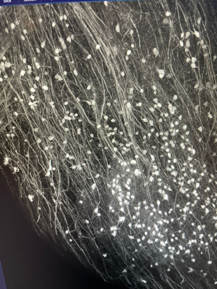
Image preprocessing. This is from the acquisition in my previous post. Final image will be gorgeous!


